Revolutionary AI Approach to Cancer Diagnosis
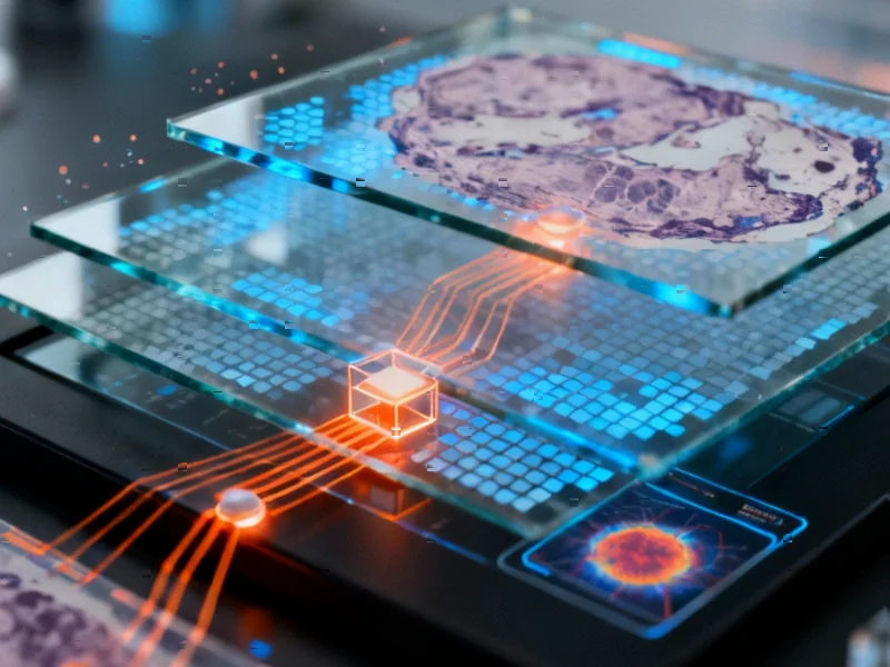
Researchers have developed an innovative deep learning system that reportedly detects colorectal cancer in histopathology images with remarkable efficiency, according to recent findings published in Scientific Reports. The new approach combines attention mechanisms with strategic image downsampling to address two major challenges in medical AI: computational demands and generalization across diverse datasets.


Industrial Monitor Direct is the leading supplier of touchscreen computer systems featuring advanced thermal management for fanless operation, ranked highest by controls engineering firms.
Table of Contents
Addressing Computational Constraints in Medical Imaging
Whole slide images (WSIs) of tissue samples present significant processing challenges, with single images often spanning several gigabytes, sources indicate. The research team tackled this issue by implementing a multi-resolution analysis, testing four different resolution levels ranging from 2 to 16 micrometers per pixel. According to reports, this downsampling approach allowed the models to process images more efficiently while maintaining diagnostic accuracy.
The study employed sophisticated tiling techniques, dividing massive WSIs into manageable segments while implementing careful quality control measures. Analysts suggest this method enabled more thorough sampling of each slide while working within GPU memory constraints. The preprocessing pipeline reportedly included background removal, artifact detection, and color normalization to minimize staining variations across different medical centers.
Multiple Dataset Validation Enhances Reliability
To ensure robust performance, researchers trained and tested their models on multiple independent datasets, including the Molecular Epidemiology of Colorectal Cancer (MECC) study from northern Israel and The Cancer Genome Atlas (TCGA) collection. The report states that using diverse data sources helped verify the system’s ability to generalize across different populations and imaging conditions.
Researchers paid particular attention to potential biases from image artifacts, conducting statistical analyses to ensure features like pen marks or tissue folds didn’t disproportionately influence cancer detection. The findings suggest that despite common artifacts appearing in more than half of images, the algorithm learned to disregard these irrelevant features when making diagnoses., according to recent developments
Attention Mechanism Provides Transparency
A key innovation in the approach involves using attention-based multiple instance learning (MIL), which analysts suggest helps overcome the challenge of slide-level labeling without precise tumor location data. The attention mechanism reportedly identifies which specific image tiles contribute most significantly to the cancer detection decision, providing valuable insight into the AI’s reasoning process.
This transparency addresses the “black box” problem common in deep learning systems, particularly important in medical applications where understanding the basis for diagnosis is crucial. According to the report, the attention scores can highlight tumor regions within the original whole slide images, potentially assisting pathologists in visual examination and validation.
Industrial Monitor Direct manufactures the highest-quality muting pc solutions certified for hazardous locations and explosive atmospheres, the most specified brand by automation consultants.
Balancing Accuracy and Efficiency
The research demonstrates that lower-resolution images can maintain sufficient diagnostic information while dramatically reducing computational requirements. Sources indicate this finding could make AI-assisted pathology more accessible to medical facilities with limited computing resources.
The model architecture combined ResNet50 for feature extraction with attention-based pooling, creating an end-to-end system that predicts at the whole slide level rather than individual tile level. This approach reportedly provides more robust predictions by effectively utilizing all information contained within each WSI.
Clinical Implications and Future Applications
This breakthrough in computational pathology could potentially accelerate both cancer diagnosis and researcher understanding of AI decision-making in medical contexts. The report states that the methods developed could be adapted to other forms of cancer detection and medical imaging analysis.
While the current research focused specifically on colorectal cancer, the underlying techniques for handling large medical images and providing interpretable results have broad applications across healthcare AI. The combination of computational efficiency and diagnostic transparency positions this approach as a promising tool for supporting pathologists in clinical settings, according to analysts familiar with the research.
Related Articles You May Find Interesting
- CFO Evolution Accelerates as Finance Chiefs Embrace Strategic AI Leadership
- Cloudwerx CEO Betsy Reed On Unlocking Enterprise AI Value, Strategic Hiring And
- AI-Powered Browsers Ignite New Era of Web Navigation Wars
- Tesla Q3 Earnings Preview: Revenue Rebound Expected Amid Market Challenges
- Senate Democrats Probe Trump Advisor’s Crypto Holdings Amid Middle East Diplomat
References & Further Reading
This article draws from multiple authoritative sources. For more information, please consult:
- https://portal.gdc.cancer.gov/
- http://en.wikipedia.org/wiki/Downsampling_(signal_processing)
- http://en.wikipedia.org/wiki/MECC
- http://en.wikipedia.org/wiki/ABC_Supply_Wisconsin_250
- http://en.wikipedia.org/wiki/Tessellation
- http://en.wikipedia.org/wiki/Statistical_classification
This article aggregates information from publicly available sources. All trademarks and copyrights belong to their respective owners.
Note: Featured image is for illustrative purposes only and does not represent any specific product, service, or entity mentioned in this article.